How AI is Changing Brain Tumor Surgery
High-tech imaging approaches allow neurosurgeons to interpret tissue samples during surgery to remove previously invisible malignant cells
By Alejandra Canales
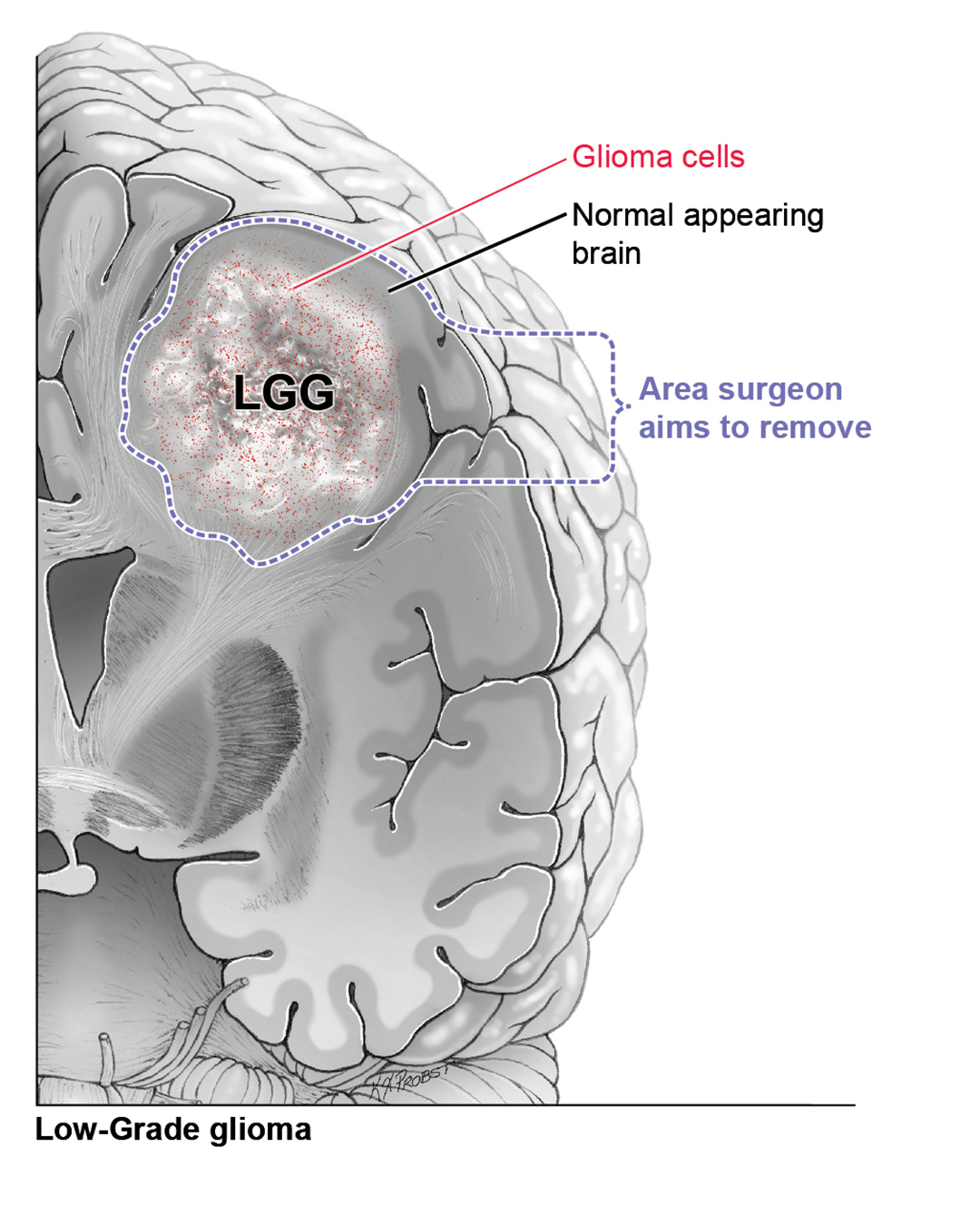
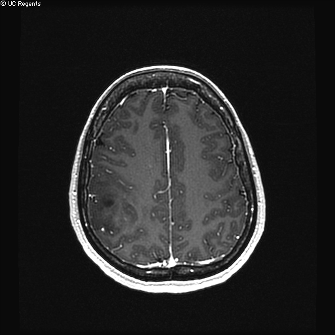
Safely removing as much of the tumor as possible is crucial to extending the survival of patients with both low- and high-grade gliomas.
This often means taking out tissue beyond the visible borders of the tumor. Although the tissue may appear normal, it typically contains tumor cells we can’t see.
But how much of the adjacent normal-appearing brain does a surgeon need to remove?
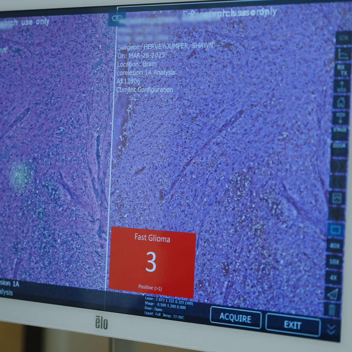
Researchers at UC San Francisco and the University of Michigan have now developed a new intraoperative tool called FastGlioma that uses AI to help neurosurgeons see in real-time where tumor cells have infiltrated brain tissue, making surgery more precise.
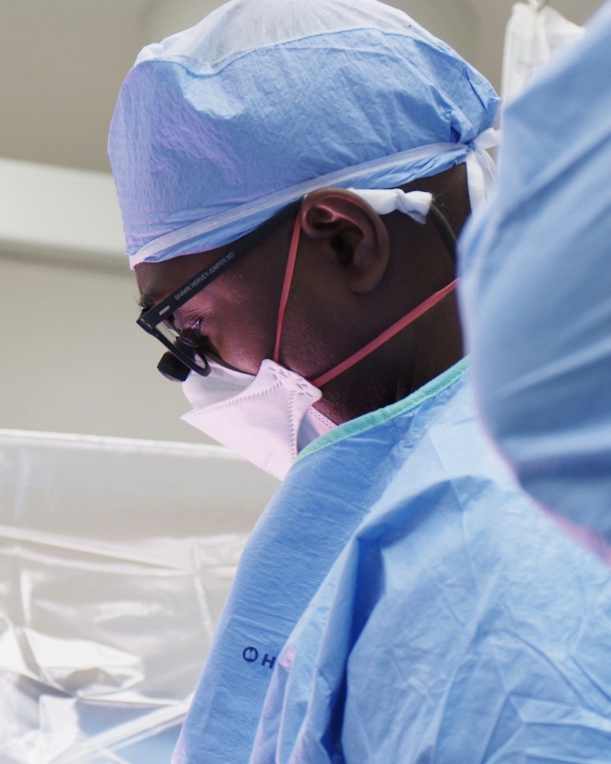
“This technology totally changes how you go into surgery,” said Shawn Hervey-Jumper, MD, the Mitchel S. Berger Endowed Professor of Neurosurgery and one of the senior co-authors of the research.
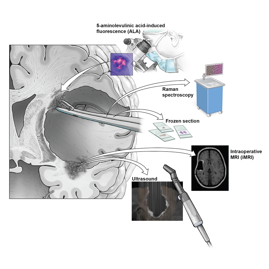
In a study published last year in Nature, the team showed that FastGlioma distinguished tumor from normal brain tumor more accurately than other approaches commonly used during surgery like 5-ALA and intraoperative MRI.
“With this new technology, we can deliver safer, more complete tumor resections,” Hervey-Jumper said.