Program Project Grant
The Program Project Grant at the UCSF Brain Tumor Center is in its tenth cycle of funding from the NIH. The Center’s first Program Project Grant was awarded in 1979 to study the biology and therapy of malignant brain tumors. Since then, the program has evolved to focus on developing and applying novel noninvasive neuroimaging techniques to clinical problems in neuro-oncology.
In the last decade, delineation of histopathological and molecular subgroups of glioma has revolutionized the field of neuro-oncology by improving diagnosis and prognosis. Research from the UCSF Brain Tumor Center also recently uncovered three distinct groups of meningiomas that can distinguish clinical outcomes, biological drivers, and therapeutic vulnerabilities from their DNA methylation patterns.
Interrogating metabolic imaging and genomic signatures of glioblastoma and meningioma will be critical to developing and evaluating new treatment approaches. For the next four years, the program will focus on four projects aimed at improving the management of patients with these tumors.
Project 1: Spatial imaging of whole tumor genomics to understand glioblastoma formation and evolution
Project Leader: Joseph Costello, PhD
This project will use image-guided whole tumor sampling and genomics to generate 3D models of individuals’ tumors from a cohort of 25 patients with newly diagnosed glioblastoma and 25 patients with recurrent glioblastoma. This approach will provide a better understanding of the genetic events that cause the tumors to form and help identify new potential therapeutic targets present throughout the tumor.
Project 2: Biological mechanisms and therapeutic vulnerability in meningioma evolution and heterogeneity
Project Leader: David Raleigh, MD, PhD
Some meningiomas can be effectively treated with surgery and postoperative radiotherapy, but others are resistant to these treatments. Project 2 aims to identify biological drivers, imaging features, and therapeutic vulnerabilities underlying different molecular subtypes of meningiomas.
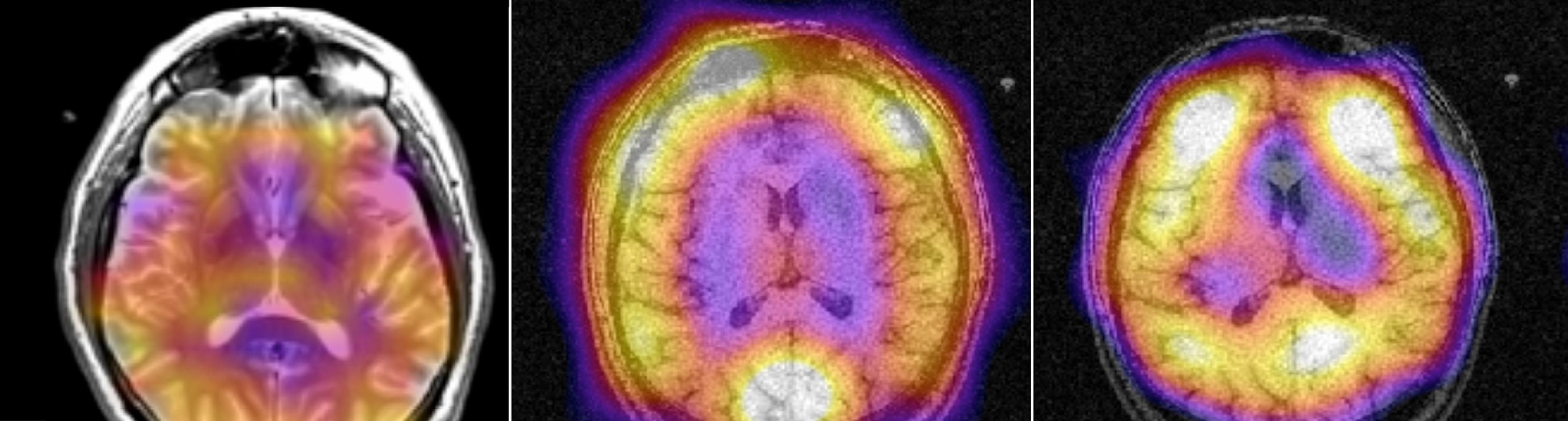
Project 3: Preclinical metabolic imaging of molecular alterations in meningiomas
Project Leader: Pavithra Viswanath, PhD
This project will use 1H and hyperpolarized 13C MR spectroscopy in preclinical models to identify different molecular subgroups of meningiomas based on the downstream changes those genetic alterations cause to their metabolism. This approach could provide physicians with a noninvasive method to assess the likelihood of an individual’s tumor responding to treatment or recurring.
Project 4: Clinical translation and validation of metabolic probes to evaluate brain tumors
Project Leader: Yan Li, PhD
Hyperpolarized 13C metabolic imaging techniques can assess dynamic changes to a tumor’s metabolism. Project 4 will use this imaging approach to evaluate early metabolic changes in patients with recurrent glioblastoma who are receiving novel treatments. This project will also develop a new hyperpolarized imaging probe to distinguish between different molecular subgroups of meningioma to aid in clinical decision making and improve patient care.