
How Neuronal Activity Reshapes the Immune Response in Brain Tumors
Antiepileptic drug generates a more antitumor immune response in mice
By studying how deadly brain tumors interact with their surrounding environment, cancer neuroscience researchers are looking to find new strategies to halt their growth.
Now a new study led by scientists at UC San Francisco reveals a three-way interaction between neurons, tumor growth, and the immune cells in the tumor microenvironment of glioblastomas.
The findings, recently published in Nature Communications, suggest that modulating neuronal activity could also be a potential approach for improving the efficacy of immunotherapies against brain tumors.
“We’ve known about the role that neuronal signaling can play in peripheral settings, like the gastrointestinal system,” said Hideho Okada, MD, PhD, a UCSF Brain Tumor Center principal investigator and the study’s co-senior author. “But this is a paradigm shift in terms of our understanding of the immune response in the brain,”
In 2023, researchers led by led by UCSF neurosurgeon Shawn Hervey-Jumper, MD, and neuroscientist Saritha Krishna, PhD showed that glioblastomas can grow more aggressively due to the connections, or synapses, that these malignant brain tumors form with neurons. More functional connectivity between the neurons and the tumor was associated with worse survival outcomes for patients.
The Hervey-Jumper lab then sought out the Okada’s lab expertise to explore the immune cell components of the tumor microenvironment. The team analyzed single-cell RNA sequencing data from patients with glioblastoma, looking for differences in the gene expression patterns between high functional connectivity (HFC) regions versus low functional connectivity (LFC) regions.
“HFC regions – more aggressive tumor regions – were correlated with more prominent immunosuppression,” said Takahide Nejo, PhD, an assistant professional researcher in the Okada lab and the study’s first author. “That was completely unexpected but perhaps the most exciting discovery in this project.”
Nejo and his colleagues found that these areas in tissue samples from glioblastoma patients had a higher proportion of anti-inflammatory tumor-associated macrophages – one of the main types of immune cells in glioblastoma.
“Tumor-associated macrophages contribute to making the microenvironment in glioblastoma more immunologically cold,” Nejo said. “They promote tumor growth and invasion and prevent antitumor immunity by suppressing T cell functions.”
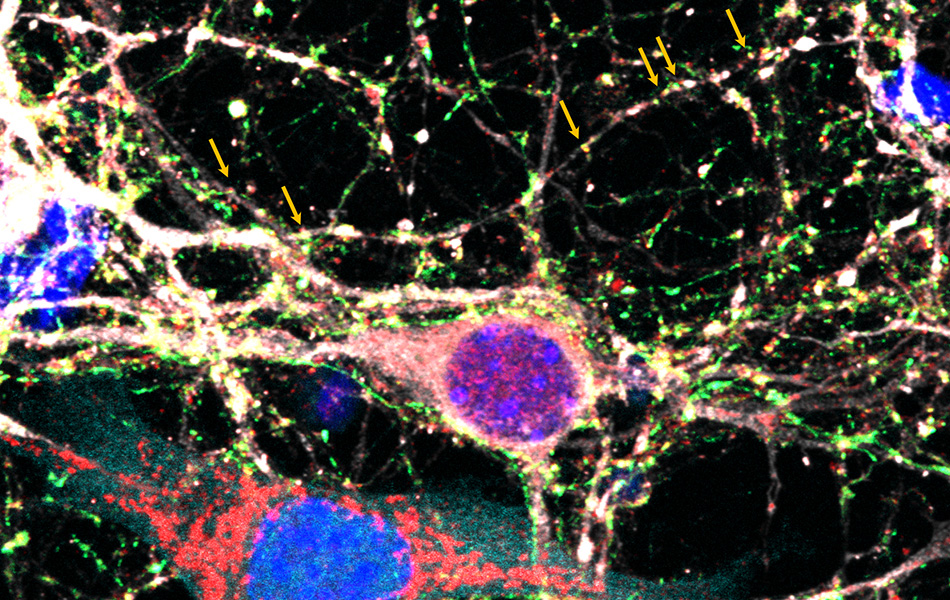
Corroborating the results from the prior 2023 study, the scientists then showed that the protein thrombospondin-1 (TSP-1) plays a key role in regulating neuronal activity between the tumor cells and the neurons through the growth of new synapses. In a mouse glioma cell line that had been genetically modified to remove TSP-1, synaptic formation and glutamatergic excitatory signals were significantly reduced.
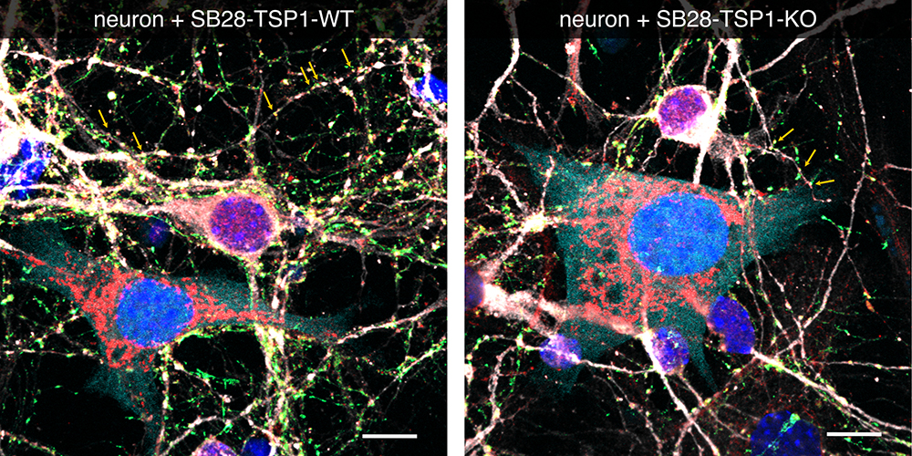
Nejo and his colleagues collaborated with the lab of Cathryn Cadwell, MD, PhD, an assistant professor of neurosurgery and pathology, to further demonstrate that TSP-1 facilitates more excitatory activity in the tumor-surrounding areas.
Without TSP-1, the glioblastoma tumor microenvironment had more antitumor macrophages and T cells.
The scientists also then showed they could replicate this effect in a mouse model of glioblastoma by inhibiting neuronal activity with an FDA-approved drug used to treat epilepsy called perampanel. The mice treated with the drug had more proinflammatory immune cell populations and generally tended to live slightly longer.
This multidisciplinary work, Nejo says, represents one of the pioneering studies investigating the very complicated crosstalk between brain tumor cells, neuronal systems, and immune components.
“We believe that by modulating the tumor microenvironment to be less immunosuppressive we can make CAR T-cell therapy more effective against brain tumors,” Nejo said. “Manipulating neural activity in the tumor microenvironment could be one of those strategies.”
Reference: Nejo, T., Krishna, S., Yamamichi, A., Lakshmanachetty, S., Jimenez, C., Lee, K. Y., Baker, D. L., Young, J. S., Chen, T., Phyu, S. S. S., Phung, L., Gallus, M., Maldonado, G. C., Okada, K., Ogino, H., Watchmaker, P. B., Diebold, D., Choudhury, A., Daniel, A. G. S., Cadwell, C. R., … Okada, H. (2025). Glioma-neuronal circuit remodeling induces regional immunosuppression. Nature communications, 16(1), 4770.