
A New Niche for Glioblastoma Immune Cells
UCSF Brain Tumor Center study finds immune cells from skull bone marrow can mediate an antitumor immune response
When Meeki Lad arrived at UC San Francisco for his away research rotation before the start of his fourth year of medical school, he didn’t have an extensive background as scientist working in a lab.
But what drew him to neurosurgery, and science more broadly, was the idea that a better understanding of the basic biology of the brain tumor microenvironment could help uncover new potential therapeutic targets.
His work in the laboratory of Manish Aghi, MD, PhD, a professor in the Department of Neurological Surgery, shows that in glioblastomas a subset of immune cells called neutrophils can flag the pathogens or cancer cells that the immune system’s T cells need to attack. Dendritic cells are usually the immune cells that carry out this process – known as antigen presentation.
The findings, published September 9 in Cancer Cell, suggest that these neutrophils could be playing an important, previously unappreciated role in mounting a response against the tumor.
Neutrophils serve as the immune system’s first line of defense. Since neutrophils are quickly replaced by other immune cells, the scientific consensus was that they wouldn’t be present within the tumor long enough to have an active part in an immune response.
“But these neutrophils in the tumor have a much longer lifespan and upregulate genes that we don't typically see in the neutrophils that circulate in the blood,” Aghi, the study’s senior author, said. “If they’re upregulating totally different genes, then that makes them totally different cells.”
Led by Lad, Aghi’s team set out to figure out what was making the tumor neutrophils behave differently.
When Lad removed these neutrophils from mice to his surprise, he found that the mice’s tumors grew larger, and they died sooner.
“This result contradicted all the prior work because it was suggesting that the neutrophils were actually suppressing tumor growth,” Lad said.
Lad then began trying to get blood neutrophils from patients to express the same genes that were present in the tumor neutrophils – a task which turned out to be impossible.
“I was just unsuccessful every time,” he said, adding that after more than a dozen failed experiments, he was forced to accept that the blood neutrophils could not be converted into tumor neutrophils.
While watching Aghi performing a craniotomy in the operating room, Lad suddenly had the idea that the tumor neutrophils might be coming from the skull bone marrow instead of the long leg bone marrow the way most neutrophils do.
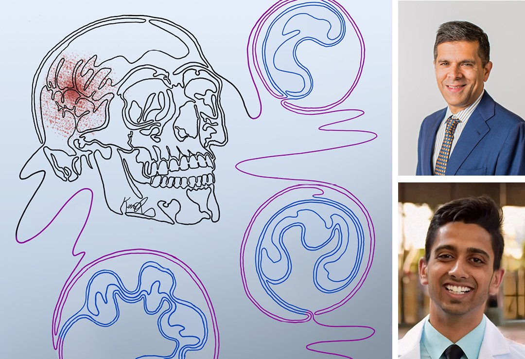
Lad examined the tumor neutrophils from both patients and mice under the microscope and saw proteins on their cell surface corresponding to markers of immature, or neutrophil precursor, cells. Since the leg bone marrow doesn’t release immature cells into circulation the researchers realized that the cells had to be coming from a source of bone marrow much closer to the tumor.
Lad and his colleagues then showed that in mice these immature neutrophils migrate from the skull bone marrow to the meninges and then into the brain or to the lymph nodes in the head and neck. Aghi says that this pathway constantly exposes the neutrophils to brain antigens, which should make them more efficient at recognizing inflammation in the brain.
Giving mice a drug called AMD 3100, which is approved for bone marrow transplants in patients with blood cancers, caused the skull bone marrow to release more of these immature cells. The mice that received this drug also lived longer, but the researchers didn’t see that effect when they depleted the mice of neutrophils before administering the drug.
Aghi’s lab is further exploring how to enhance the tumor neutrophils’ effect on the immune response. Although dendritic cells can also stimulate an immune response, Aghi says that neutrophils seem to be more efficient as antigen presenting cells and are more abundant in the tumor. These factors might make an approach combining traditional immunotherapies that activate T cells with AMD 3100 more promising.
The researchers are also interested in how skull bone marrow and all the immune cells it generates interact with the tumor.
And for Lad, this project in the Aghi lab made rethink his career trajectory as he discovered how much he liked doing research. Now, having switched onto an MD-PhD track, he’s continuing these studies as part of his doctoral thesis.
Reference: Lad M et al. Glioblastoma induces the recruitment and differentiation of dendritic-like “hybrid” neutrophils from skull bone marrow. Cancer Cell. 2024 Sep 9; 42(9):p1549-1569.e16.